Cardiac CT can double as BMD screening for osteoporosis
17 Jul 2020
byPearl Toh
Very low thoracic bone mineral density (BMD) measured on cardiac CT scan is predictive of a higher fracture risk, especially osteoporosis-related fracture — thus highlighting the potential utility of cardiac CT for opportunistic BMD screening.
“Many osteoporotic fractures occur in individuals who have never been screened,” wrote Professor Miriam Bredella of Harvard Medical School, Boston, Massachusetts, US, in a linked editorial. [Radiology 2020;doi:10.1148/radiol.2020202374]
“Effective anti-osteoporotic treatment exists and so, identifying individuals with greater fracture rate who may benefit from such treatment is imperative,” said lead author Dr Josephine Therkildsen from Herning Hospital, Hospital Unit West in Herning, Denmark.
“The prevalence of chest CT imaging provides a unique opportunity for opportunistic BMD testing,” Therkildsen and co-authors pointed out. “Routine cardiac CT can be used to help measure thoracic BMD to identify individuals who have low BMD and a greater fracture rate.”
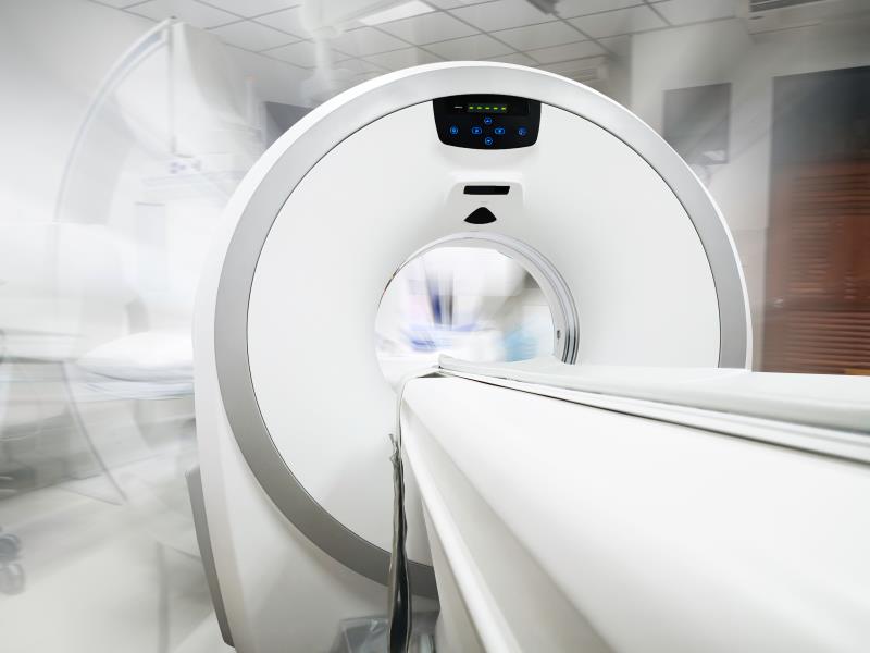
As cardiac CT also images the thoracic vertebrae (in addition to the heart), this can be exploited for measurement of spine BMD.
The prospective cohort study involved 1,487 consecutive participants who were referred for cardiac CT to assess ischaemic heart disease. During a median follow-up of 3.1 years, 80 participants (5.3 percent) had an incident fracture. Among the 80 incident fractures, 31 were osteoporosis related. [Radiology 2020;doi:10.1148/radiol.2020192706]
The risk of any fracture was significantly higher in participants with very low BMD on cardiac CT (<80 mg/cm3) compared with those with a normal BMD (>120 mg/cm3; hazard ratio [HR], 2.6; p=0.002).
In particular, very low BMD was associated with a substantially elevated risk of any osteoporosis-related fracture vs normal BMD (HR, 8.1; p=0.001).
The associations with very low BMD remained for any fracture (HR, 2.1; 95 percent confidence interval [CI], 1.1– 4.2) and any osteoporosis-related fracture (HR, 4.0; 95 percent CI, 1.1–14.6) after adjustment for age and sex.
While current guidelines recommend using DEXA* as a diagnostic test for osteoporosis, a previous study has shown that quantitative CT better predicts vertebral fractures than DEXA scan, the authors noted. [Bone 2016;92:100-106]
“Our results represent a step toward appraisal and recognition of the clinical utility of opportunistic BMD screening from cardiac CT,” said Therkildsen and co-authors.
According to Therkildsen, it is relatively easy to add BMD testing to cardiac CT as it does not require additional time and radiation exposure to the patient. BMD measurements are possible using existing CT images as long as there are continuous scanner stability, systematic imaging acquisition, plus a suitable calibration system.
“We believe that opportunistic BMD testing using routine CT scans can be done with little change to normal clinical practice and with the benefit of identifying individuals with a greater fracture rate,” said Therkildsen.
Future studies will help determine the optimal thresholds for treatment based on quantitative CT measurements of thoracic spine BMD, the researchers suggested.