The robot will examine you now: Remote ultrasound of COVID-19 patients a success
29 Jul 2020

Jairia Dela Cruz
Jairia Dela Cruz
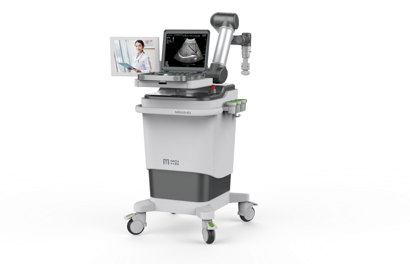 The patient-side subsystem of the MGIUS-R3. Image provided by Ruizhong Ye, MD, Department of Ultrasound Medicine, Zhejiang Provincial People's Hospital & People's Hospital of Hangzhou Medical College.
A robotic device controlled remotely via the internet has successfully performed chest ultrasound of patients with COVID-19, obtaining characteristics necessary for cardiopulmonary assessment, as shown in a study.
Named MGIUS-R3, the robotic ultrasound consisted of two paired subsystems, each located at a different hospital, connected through a 5G network. The patient-side subsystem comprised a robotic arm fitted with an ultrasound imaging that used a 1.0‒5.5 MHz convex array probe. This arm was remotely operated by sonographers.
The device successfully completed cardiopulmonary assessment for all patients included in the study, with each examination taking 10‒20 minutes on average, and there was no obvious delay in scanning. MGIUS-R3 yielded ultrasound image information, such as distribution characteristics, morphology of the lungs and surrounding tissue lesions, left ventricular ejection fraction (LVEF), pericardial and pleural effusion, and lung ultrasound score (LUS). There were no reports of significant examination-related complications.
In the images, severe COVID-19 patients, compared with those whose disease was nonsevere, showed significantly more diseased regions in the lungs (median, 6.0 vs 1.0; p<0.05) and had higher LUS (median, 12.0 vs 2.0; p<0.05). Pleural effusions were seen in one nonsevere patient (5-mm deep; 8.3 percent) and three severe patients (14-mm deep; 27.3 percent), with the pleural line described as thick and rough. [Chest 2020;doi:10.1016/j.chest.2020.06.068]
Finally, four cases of pericardial effusion (3–10 mm) were found, all of which were detected in the severe group (36.4 percent vs 0 percent; p<0.05). The LVEF and ventricular area ratio of the heart were normal in all patients.
In total, 23 COVID-19 patients had undergone an ultrasound examination using MGIUS-R3; 11 had severe disease and 12 had nonsevere disease (mean age, 70.6 years vs 56.6, years respectively; mean oxygen saturation, 93.0 percent vs 99.8 percent). An assistant stood by during the procedure to help disinfect the instrument, apply medical coupling agents, and adjust the position of severely ill patients appropriately.
The present data suggest that “ultrasonography might be helpful for evaluating COVID-19 severity, and the 5G-based robot-assisted remote ultrasound system could achieve the same effect as a face-to-face, close-range ultrasound examination,” according to investigators from China.
COVID-19 is diagnosed using high-resolution computed tomography (CT) most of the time. This approach boasts high spatial resolution and multiplanar and multidirectional display of lesion details, but at the same time, it exposes patients to ionizing radiations. Moreover, patients need to be transported to the scanner, and doing this for those with severe disease is risky. Plus, disinfecting the scanner takes a lot of work. [J Am Assoc Nurse Pract 2016;28:659-667; Am J Roentgenol 2013;201:W81-87]
In comparison, ultrasonography can be performed bedside, offering greater convenience and dynamics. It facilitates the diagnosis of lung diseases, rapid confirmation of acute respiratory failure causes, and qualitative assessment of pleural effusion (free or wrapped), among others. [J Ultrasound Med 2020;doi:10.1002/jum.15285. 2020]
“Although the initial application of the 5G-based robot-assisted remote ultrasound system in the COVID-19 epidemic has achieved good results, it cannot wholly replace CT and other examinations,” the investigators acknowledged.
They believe that the integration of artificial intelligence (AI) should greatly increase the scope of use for robot-assisted remote ultrasound systems, saying that it would “facilitate the diagnosis of lung lesions objectively and accurately, and implement automatic switching between probes on the ultrasonic robot system to [enable] optimal imaging of multiple organs and improve image quality.” [JMIR Med Inform 2019;7:e10010; Thyroid 2017;27:546-552; Med Phys 2017;44:1678-1691]