High fructose intake not linked to negative metabolic effects in healthy individuals
10 Feb 2020
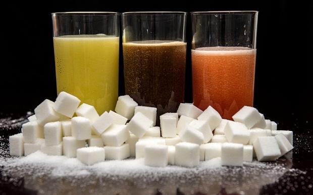
Prolonged ingestion of a high-dose dietary fructose does not result in relevant metabolic consequences in the presence of a stable energy intake, slightly lower body weight and potentially incomplete absorption of orally administered fructose load, according to a study.
This indicates that “young, metabolically healthy [individuals] can at least temporarily compensate for increased fructose intake,” the investigators said.
High intake of fructose led to reduced consumption of other dietary sugars and did not increase total daily energy intake. There was also no change in ectopic lipid deposition and postprandial glycogen storage in the liver and skeletal muscle.
Postprandial changes were noted in hepatic lipids (Δhepatocellular lipid [HCL]_healthy_baseline vs follow-up: −15.9±10.7 vs −6.9±4.6; p=0.17) and hepatic glycogen (Δglycogen_baseline vs follow-up: 64.4±14.1 vs 51.1±9.8; p=0.42). Myocardial function and myocardial mass remained stable.
Not surprisingly, insulin-resistant patients with nonalcoholic fatty liver disease (NAFLD) were shown to have impaired hepatic glycogen storage and increased ectopic lipid storage in the liver and skeletal muscle.
This study included 10 healthy individuals (aged 28±19 years; body mass index [BMI], 22.2±0.7 kg/m2) who underwent comprehensive metabolic phenotyping prior to and 8 weeks following a high-fructose diet (150 g daily), as well as 11 patients with NAFLD (aged 39±3.95 years; BMI, 28.4±1.25 kg/m2) as positive controls.
The investigators employed a 2-step hyperinsulinaemic euglycaemic clamp to analyse insulin sensitivity, as well as evaluated postprandial interorgan crosstalk of lipid and glucose metabolism by determining postprandial hepatic and intramyocellular lipid and glycogen accumulation using magnetic resonance spectroscopy (MRS) at 7 T. They also assessed myocardial lipid content and myocardial function via 1H MRS imaging and MRI at 3 T.