Novel PET-CT method superior to conventional imaging in detecting high-risk localized prostate cancer
31 Mar 2020
byPank Jit Sin
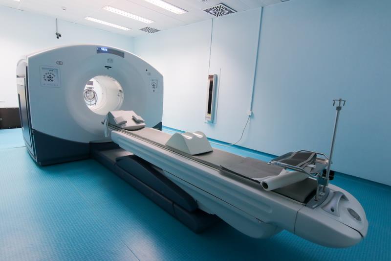
The multicenter, two-arm, randomized study looked at men with biopsy-proven prostate cancer and high-risk features at 10 hospitals in Australia. [Australian New Zealand Clinical Trials Registry ANZCTR12617000005358] Patients were randomized to undergo either conventional CT and bone scanning or gallium-68 PSMA-11 PET-CT. First-line imaging was performed within 21 days of randomization. Patients were crossed over unless three or more distant metastases were identified. The study’s primary outcome looked at the accuracy of first-line imaging for identifying either pelvic nodal or distant-metastatic diseases, which is defined by the receiver-operating curve using a predefined reference standard, and includes histopathology, imaging and biochemistry at 6-month follow-up.
The study found that PSMA PET-CT had a 27 percent (95 percent CI 23–31) greater accuracy compared to conventional imaging (92 percent [88–95] vs 65 percent [60–69]; p<0.0001). Additionally, conventional imaging was less sensitive and specific compared to PSMA PET-CT. PSMA PET-CT also proved to be superior to conventional imaging for patients with pelvic nodal metastases and patients with distant metastases.
The study also noted that radiation exposure was higher in conventional imaging compared with PSMA PET-CT (19.2 mSv vs 8.4 mSv; p<0.001).