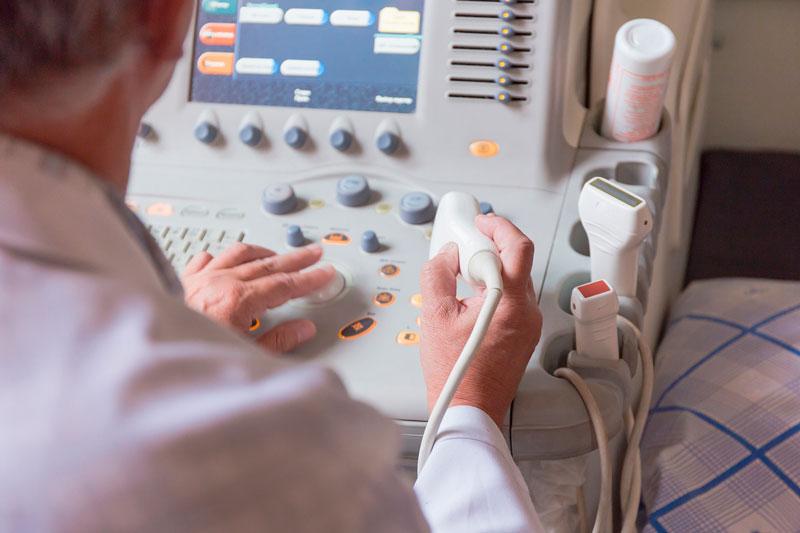
Transperineal ultrasound useful for predicting endoscopic, histological healing in ulcerative colitis


Bowel wall thickness in transperineal ultrasound correlates well with both endoscopic severity and histological activity in patients with ulcerative colitis (UC), as shown in a study. The combination of transperineal and transabdominal ultrasound enables visualization of the entire colorectum and is an ideal modality to monitor disease course through a treat‐to‐target strategy.
“UC is a diffuse nonspecific inflammatory disorder of the large bowel presenting with superficial erosions and ulceration… Symptoms and endoscopic findings do not always correlate, thus, it is necessary to frequently monitor the endoscopic activity and optimize the treatment,” according to the investigators. [J Crohns Colitis 2016;10:1303-1309]
“Transabdominal ultrasound has been reported to well correlate with the endoscopic severity of UC while it can visualize the extent and severity with very high patients' acceptability… However, it is also known that rectal inflammation is difficult to evaluate because of the distance from the abdominal wall,” they continued. [J Crohns Colitis 2018;12:1385-1391; J Gastroenterol 2019;54:521-529; J Crohns Colitis 2019;13:S218]
In the current study, the investigators evaluated the utility of transperineal ultrasound in terms of predicting endoscopic and histological findings of the rectum. They examined 53 consecutive patients who underwent transperineal in combination with transabdominal ultrasound within a week before or after colonoscopy with rectal biopsy. Endoscopic healing was defined as Mayo endoscopic subscore (MES) ≤1, while histological healing was defined as Geboes score <2.1, Robarts histopathology index ≤6 and Nancy index ≤1.
Colonoscopy findings showed excellent correlation with those of transabdominal ultrasound in all segments except for the rectum. Meanwhile, rectal MES and histological indices correlated well with rectal bowel wall thickness and Limberg score in transperineal ultrasound. [Aliment Pharmacol Ther 2020;doi:10.1111/apt.15767]
Bowel wall thickness ≥4 mm predicted MES with greater accuracy in transperineal than in transabdominal ultrasound (area under the curve [AUC], 0.904 vs 0.667; p=0.0002), with respective sensitivity of 100 percent vs 96.3 percent and specificity of 45.8 percent vs 12.5 percent. The predictive value of Limberg score was also improved by transperineal ultrasound.
Likewise, bowel wall thickness and Limberg score in transperineal ultrasound were able to predict activity in Geboes score, Robarts histopathology index and Nancy histological index (AUCs, 0.81–0.89).
In multivariable logistic regression models, bowel wall thickness in transperineal ultrasound was the sole predictor of rectal endoscopic (MES >1 in rectum: adjusted odds ratio [aOR],3.18; p=0.0003) and histologic healing (Geboes score ≥2.1: aOR, 2.73; Robarts histopathology index >6: aOR, 3.27; Nancy histological index >1: aOR, 2.60; p<0.05). Its predictive ability was better than that of faecal calprotectin.
“Our study identified transperineal ultrasound as an extremely useful additional examination to [and] that compensates for the weakness of transabdominal ultrasound,” according to the investigators. “Transperineal ultrasound is patient‐friendly and does not require preparations such as fluids, laxatives and fasting. It can also visualize the extent and distribution of lesions which cannot be obtained through biomarkers.” [Clin Gastroenterol Hepatol 2018;16:1089-1097.e4]
While faecal calprotectin may be used to screen rectal inflammation when transabdominal ultrasound do not show the activity in the colon, as shown in a recent study, performing transperineal ultrasound in addition to transabdominal ultrasound on site is much more convenient compared with this two‐step strategy, they pointed out. [J Crohns Colitis 2018;12:1385-1391]
“Taken together, these findings may suggest the exclusive usefulness of ultrasound as a cross-sectional imaging in assessing subepithelial inflammation which cannot be accurately assessed by endoscopy. This may allow us to predict future progress of bowel damage in UC patients,” the investigators said. “Further study is needed to confirm the potential usefulness of ultrasound, especially transperineal ultrasound.”