Chromoendoscopy detects more Lynch syndrome lesions than white light endoscopy
22 Nov 2019
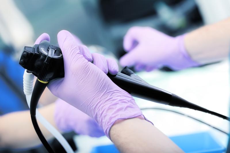
In patients with Lynch syndrome, chromoendoscopy (ChE) allows for the detection of more lesions than white light endoscopy (WLE), especially in terms of adenomas, flat lesions and proximal lesions, reports a recent meta-analysis.
Researchers retrieved four studies from Medline, which compared the detection of neoplastic lesions using between least two endoscopic imaging methods. This resulted in a sample of 164 patients undergoing ChE and 190 undergoing WLE. Only prospective studies with parallel or crossover designs, and which included Lynch syndrome patients, were eligible for inclusion.
A total of 311 and 169 lesions were detected by ChE and WLE, respectively. The resulting detection rate of ChE was almost twice that of WLE (rate ratio [RR], 1.97, 95 percent confidence intervals [CI], 1.63–2.38). ChE was also able to detect more adenomas than WLE (81 vs 56), such that its detection rate was at least 1.5 times greater (RR, 1.53, 95 percent CI, 1.07–2.17). More than half of all the adenomas in all studies included was detected by ChE.
Flat lesions were also more detectable using ChE than WLE (173 vs 52; RR, 3.4, 95 percent CI, 2.47–4.67). More than three-fourths (77 percent) of all flat lesions were identified using ChE. A similar effect was reported for proximal lesions, which were detected by ChE at almost three times the rate of WLE (86 vs 30; RR, 2.93, 95 percent CI, 1.91–4.50).
“More studies are needed comparing [high-definition] technology and advanced imaging methods with ChE for adenoma detection … and to assess the impact of ChE on advanced polyps and cancer detection,” said researchers.