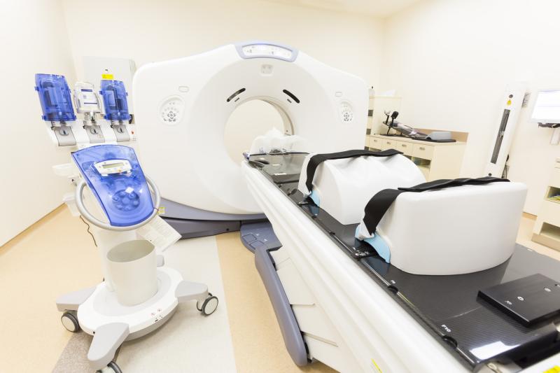
Imaging modality appears to significantly affect the Medina classification, a recent study has found. Coronary computed tomography angiography (CTA) provides greater reproducibility of results compared with invasive coronary angiography (ICA).
The study included 363 patients with 400 bifurcation lesions. Twenty-six (mean age, 63.6±11.0 years; 73 percent male) had side branch (SB) occlusions, while the remaining 337 (mean age, 64.3±9.9 years; 73 percent male) did not.
The distribution of Medina classifications was significantly different between modality groups (p<0.001). This was driven primarily by a much lower prevalence of SB disease in the CTA arm. True bifurcations were also significantly less prevalent in the CTA group (p<0.001). The resulting agreement between CTA and ICA for Medina assessment was poor (kappa, 0.189).
Similarly, CTA and ICA were also poorly in sync when it came to the identification of true bifurcations (kappa, 0.199), and bifurcations with any lesions involving proximal main vessel and SB (kappa, 0.172). Out of 400 bifurcations, 63.3 percent of the classifications delivered were discordant.
Using a subset of 100 lesions, the reproducibility of Medina classifications was compared between imaging modalities. Interobserver agreement upon CTA was moderate for Medina (kappa, 0.557) and true bifurcations (kappa, 0.536). On the other hand, ICA yielded worse estimates, with only fair agreement for Medina (kappa, 0.346) and true bifurcations (kappa, 0.323).
“[T]he present data underscore the higher level of reliability and certainty with regard to noninvasive evaluation of bifurcation lesions as compared with invasive angiography,” said the researchers.