(JM country specific)Clues to help manage pediatric skin infections
30 Jul 2024
byDr. James Salisi
Diagnosing fungal and viral skin infections may pose a challenge to many clinicians. But according to Dr. Roberta Romero, a fellow of the Philippine Dermatological Society and officer-in-charge of the Tropical Diseases Foundation at the Makati Medical Center, there are distinct clues in the history and physical examination of a patient that can help the clinician diagnose and treat pediatric skin conditions caused by dermatophytes, yeasts and viruses.
Fungal infections
“[The] first clue is, are there predisposing factors? [I]s there trauma to the skin or constant rubbing? Is there increased moisture like sweating? [Is their warmth? Poor hygiene?]” Romero said. Also, prolonged use of antibiotics and the immunocompromised state can predispose an individual to opportunistic skin infections.
Dermatophyte infection is common. An example is tinea corporis, a type of ringworm that appears on hairless skin. It can be recognized by its red, scaly papules that spread peripherally where the slow-growing dermatophyte is most active. Papules coalesce and lesions become annular. The lesions are itchy if very inflammatory, especially in the groin area. The clues that should alert the clinician are the advancing border and slow growth.
“So look for the border. Usually … the body somehow recognizes [something] as foreign, as an invader and starts to clear the center so that fungus does one thing ahead and moves to virgin territory. If you look at the fungus, it is no longer at the center as a general rule but it has moved outward,” Romero explained.
Not all rings are ringworms. Many conditions may manifest like the ring-shaped lesions of skin fungal infections, such as urticaria, which is explosive and rapidly coalescing, with edematous and erythematous borders; and atopic dermatitis, which manifests as an annular configuration and can be distinguished by the dry skin outside of the lesion.
Other lesions that may look like fungal infections are nummular eczema, with very pruritic weeping lesions, pityriasis rosea and impetigo contagiosa. The fast rate with which these lesions appear distinguishes them from fungal
infections.
Tinea capitis is more common than the other ringworms and the clue to remember is the age at which this infection usually occurs – prepubertal age. Children usually have low sebum, which has antifungal properties. Tinea capitis manifests as patchy hair loss with scales.
Sometimes in cases of tinea capitis caused by Trichopython species, it can be very inflammatory and may manifest as papules and pustules with cervical lymphadenopathy. This infection may also manifest as “black dot” tinea capitis, where fungi get into the hair, causing it to fall off; or as inflammatory tinea capitis with pus, which tests positive for fungi, not bacteria; or as kerion, a boggy, inflammatory lesion which is found only in tinea capitis. Scarring alopecia is a complication of tinea capitis when it is not treated in a timely manner.
Tinea ungium is not common in children. It is usually associated with tinea pedis. Toenails are more commonly involved than fingernails. A clue to look for is fungal debris either under or on top of the nail.
Not all dystrophic nail changes are caused by fungus. In fact, 50 percent of nail fungus diagnoses are wrong. Twenty-nail dystrophy is one example of nail dystrophy diagnosed wrongly as fungal infection. Conditions that can be mistaken for fungal nail infection but are actually part of a medical condition include pitting nails in psoriasis, onychomadesis after Steven Johnson syndrome or after hand, foot and mouth disease, and nail disease after dengue.
Although the clinical picture of dermatophyte infection is often distinctive, there is room for laboratory tests of skin scrapings such as fungal culture and KOH smear, which will show long, branching septate hyphae.
Topical anti-fungals such as azoles and terbinafine may be used for 2 to 4 weeks. Usually the treatment is continued for 1 week after lesion clears up. Oral anti-fungals are used when the lesions are not responding to topicals or when they involve the nails and hair-bearing areas. The duration and dose of oral anti-fungal treatment varies depending on the location of the lesions and the weight of the pediatric patient, respectively.
Pityriasis versicolor and candidiasis are two of the common superficial yeast infections. The etiologic agents are lipophillic or have a propensity to grow in oily surfaces conducive to their growth.
Malassezia furfur causes pityriasis versicolor. Several factors make it pathogenic such as high temperature and humidity, hyperhidrosis, increased sebum production, steroid therapy, immunodeficiency and heredity.
The fingernail sign, the fine and branny desquamation caused by scratching, is a clue to its diagnosis. Versicolor means multicolored and this is manifested in the hypopigmented to paint pink-red to tan-dark brown skin lesions which are discrete coalescent ovoid macules found in the upper back, chest, arms, forehead and temples of patients. In infants, the area of predilection is between the eyebrows.
The KOH smear of pityriasis versicolor will reveal the “spaghetti and meatballs” appearance, which are really the hyphae and spores of P. ovale. Zinc pyrithione shampoo may be applied on the skin and left for 10 minutes daily before rinsing it off for 1 to 2 weeks. Another option is 2 percent ketoconazole shampoo 5 minutes daily for 3 days. In young adults, azoles such as itraconazole and fluconazole may be used for extensive lesions resistant to topical or for frequent relapse.
The yeast Candida is limited to skin and mucous membranes when the barrier is disrupted. Areas at risk are macerated, damaged or inflamed skin. Thrush or acute pseudomembranous candidiasis should be suspected in white to gray, cheesy-looking colonies on the mucous membranes of the mouth, which reveal a raw red base upon gentle removal. Other clues are well-demarcated erythema with peripheral scale, satellite papules or pustules, inguinal creases involvement and “beefy red” erythema.
A topical anti-candidal agent such as nystatin or azole with or without topical steroid may be used to manage candidiasis. Treatment should be continued for 3 more days after the lesions are cleared. For oral thrush, oral myscostatin or fluconazole may be used.
Viral skin infections
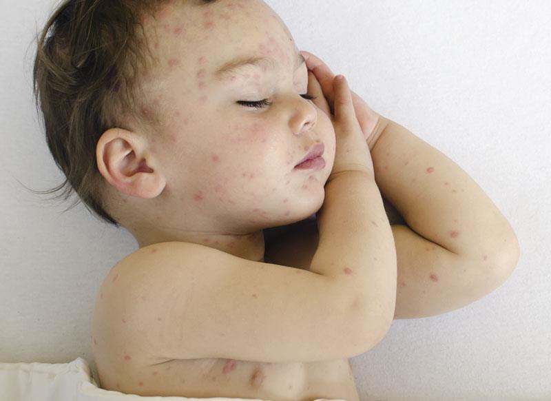
Romero explained that hand, foot and mouth disease, molluscum contagiosum and verruca vulgaris are common viral skin infections in children. When shallow grayish ulcers on erythematous base appear in the mouth, hands and feet and/or buttocks, hand foot and mouth disease should be considered. The rash usually lasts for 2 to 7 days with or without fever, sore throat, anorexia, malaise and abdominal pain. Patients are treated symptomatically.
Flesh-colored and umbilicated papules are hallmarks of molluscum contangiosum, which is very infectious. The lesions may spontaneously resolve in 6 to 9 months but for more persistent and progressive lesions, treatment includes curettage, topical cantharidin, tretinoin cream and imiquimod cream. Pediatricians should recognize, refer and avoid giving topical steroid but may try tretinoin or imiquimod.
Lastly, warts or verruca vulgaris are rough and hyperkeratotic skin lesions caused by human papillomavirus types 1, 2, 4 and 7.
The clue to diagnosing this condition is the presence of thrombosed blood vessels or black dots. Forty percent of cases spontaneously resolve after 2 years. If they are persistent or multiplying, Romero suggested cryosurgery, dessication and curettage, or chemical destruction using cantharidin plus 40 percent salicylic acid.