Hypertension alters structure of deep venous plexus
18 Jun 2020
byTristan Manalac
Hypertension decreases retinal vessel density and increases the foveal avascular zone, particularly in the deep venous plexus, according to a recent Singapore study.
“In this population-based case control study, we found that deep macular flow is decreased and the size and perimeter of the deep foveal avascular zone are increased in participants with hypertension compared to normal controls,” researchers said.
Drawing from the Singapore Chinese Eye Study, researchers conducted a case-control study of 94 eyes, from 94 patients (mean age, 64.77±9.03 years; 50 percent female) with systemic hypertension, and 46 normal eyes (46 controls; mean age, 58.3±4.62 years; 54.35 percent female). The hypertensive group had lower superficial and deep macular flow (p<0.001) upon univariate assessment. [Sci Rep 2020;10:9580]
The deep parafoveal and total flow densities, as well as the parafoveal inner retina and total parafoveal thicknesses, were likewise significantly reduced in hypertensive eyes.
Controlling for age, sex, intraocular pressure, logMAR visual acuity, spherical equivalents, and mean arterial pressure (MAP), a multivariate comparison found that macular flow in the deep layer remained the only variable significantly reduced in hypertensive vs control patients (1.21±0.1 vs 0.92±0.2; p=0.002).
On the other hand, adjusting for confounders gave rise to new between-group differences. The size of the manual foveal avascular zone in the deep layer, for instance, was significantly greater in hypertensive eyes (3.47±0.38 vs 3.15±0.3; p=0.03). The same was true for the associated perimeter of the foveal avascular zone (22.74±1.35 vs 21.4±1.1; p=0.016).
The size of the foveal avascular zone in the superficial layer, in contrast, was comparable between subgroups. Between-group differences in the deep parafoveal and total density and in the parafoveal inner and total retina thicknesses were attenuated.
“The study of deep (DVP) vs superficial (SVP) vascular plexus alterations may have important clinical implications as it may have a greater effect on the outer retinal layers, and this may affect visual functioning; however, more studies are needed to verify this relationship,” the researchers said.
“It must be considered also that in all studies examining quantitative changes in the SVP and DVP separately, accurate segmentation and removal of projection artifact is essential, and this can sometimes be challenging in eyes with significant diseases that alter the anatomical retinal layers,” they added.
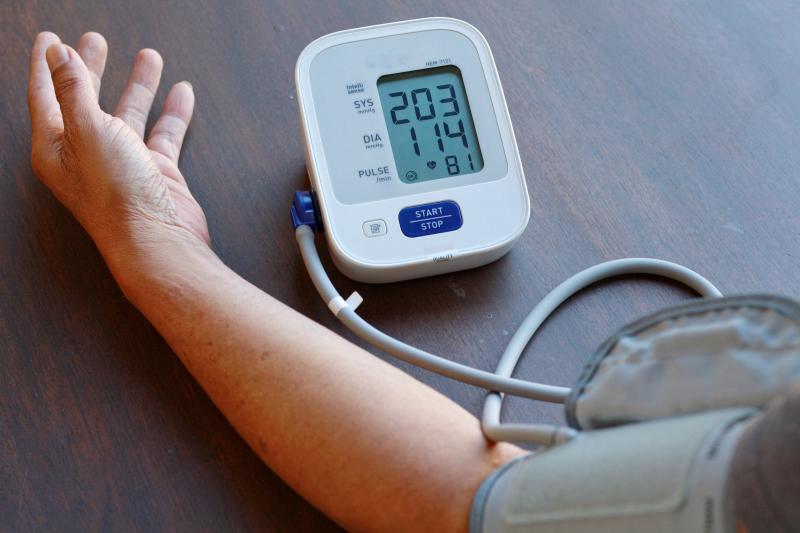
In the present study, hypertension was defined as having systolic blood pressure ≥140 mm Hg or diastolic blood pressure ≥90 mm Hg. Self-reports of diagnosis or use of medications were also considered as eligibility criteria. Optical coherence tomography angiography was used in the evaluation of the SVP and DVP.
“While we noted the number and classes of hypertensive agents, we did not do any subgroup analysis correlating type and number of medications with OCTA parameters due to insufficient numbers,” the researchers said.
“This is a possible area for future research as improvement in vascular rarefaction has been reported with the use of various antihypertensive agents, and it would be interesting to note if there were differences in improvement in rarefaction between classes of antihypertensive [agents],” they added.