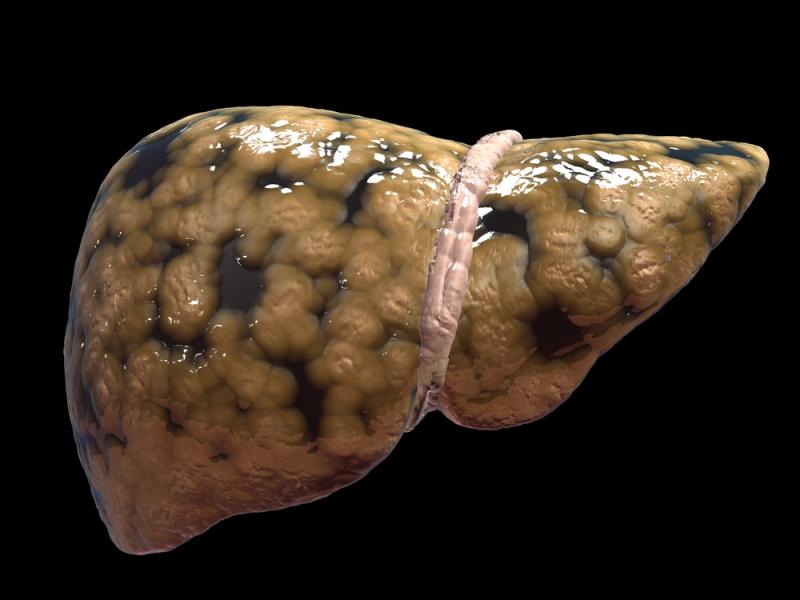
Magnetic resonance imaging may not be a reliable strategy for detecting changes in liver histology and may thus have limited value in assessing treatment response in patients with fatty liver, reports a new study.
The study included 121 patients with nonalcoholic steatohepatitis (NASH) who had paired measurements of intrahepatic triglyceride (IHTG) content, collected through magnetic resonance spectroscopy and liver histology.
Researchers found no significant correlation between changes in IHTG content and histological response to pioglitazone. For example, the resolution of NASH appeared to be comparable between those who showed a more-than 30-percent drop in IHTG content and those with a lower degree of reduction.
Similarly, a 2-point reduction in the nonalcoholic fatty liver disease activity score without worsening fibrosis occurred comparably in those who did vs did not show a 30-percent drop in IHTG. At no specific IHTG cutoff point was the prediction of NASH resolution significant.
Moreover, changes in IHTG, when plotted alongside the rate of improvement of different histological parameters, showed no significant predictive patterns.
Changes in lobular inflammation, liver fibrosis or hepatocyte ballooning also occurred independently of reductions in IHTG content.
“In the current work, we have shown that changes in IHTG content after treatment with pioglitazone or placebo do not predict histological or metabolic changes. The clinical implication of our findings is that MRI-based imaging may not be as reliable as previously believed to guide about the clinical efficacy of a given intervention in patients with NASH,” said researchers.