Brain altered in T2DM patients
04 Jul 2020
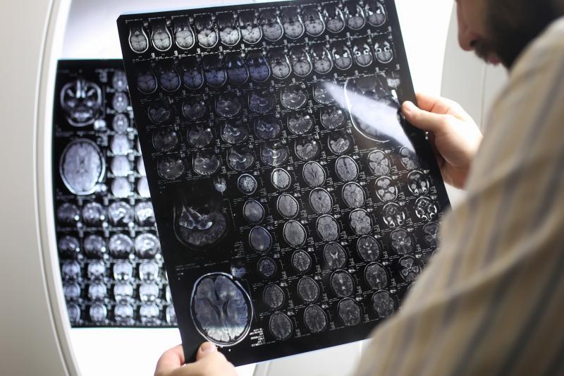 Radiologists interpret complex medical imaging to arrive at an accurate diagnosis
Patients with type 2 diabetes mellitus (T2DM) may also sustain brain damage in regions of the brain involved in cognition and emotion, a new study has found. This provides structural bases for mood and cognitive deficits in T2DM patients.
The study included 34 T2DM patients and 88 healthy controls, in whom changes in grey matter were assessed using high-resolution T1-weighted images, collected through magnetic resonance imaging. The Beck Anxiety and Depression Inventories were used to score anxiety and depression symptoms, respectively, while cognition was evaluated using the Montreal Cognitive Assessment.
Depression (p=0.001) and anxiety (p=0.003) were significantly greater in diabetic participants than in controls, while cognition was significantly worse (p=0.002). The language subdomain, in particular, saw strong impairments (p<0.001).
Several areas of the brain showed decreased regional grey matter volume in diabetics vs controls, which were particularly marked in the following: bilateral anterior and posterior insular cortices; anterior, mid, and posterior cingulate gyri; the hippocampus; the amygdala; the bilateral pre- and post-central gyrus; and the inferior, mid, and superior frontal cortex, among many other regions.
In turn, these structural changes correlated with anxiety and depression burden. For instance, anxiety shared an inverse association with grey matter volume in the bilateral caudate, bilateral mid, left superior, and right inferior frontal cortices.
Similar patterns of effect were found with depression scores, while global cognition showed positive correlations with grey matter volume in different brain regions.