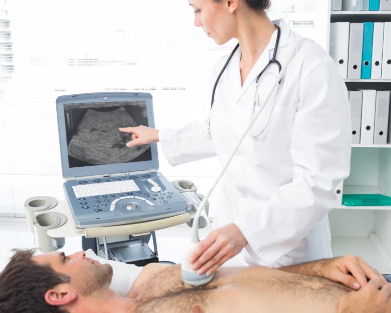
Routine use of lung ultrasound (LUS) is recommended in the diagnostic algorithm of respiratory diseases in a real-life setting, according to a multicentre prospective study.
The investigators enrolled 509 consecutive patients admitted for respiratory-related symptoms to both emergency and general medicine wards over a period of 2 years. Participants were evaluated using LUS and chest radiography (CXR).
Expert operators who were blinded to the medical history and laboratory data of patients conducted the LUS. Computed tomography (CT) of the chest was performed in case of discordance between the LUS and CXR, suspected lung cancer and an inconclusive diagnosis. A CT diagnosis was considered the gold standard.
A significant difference in sensitivity and specificity was observed between LUS and CXR as shown by receiver operating characteristic curve analyses (LUS-AUROC, 0.853; specificity, 81.6 percent; sensitivity, 93.9 percent vs CXR AUROC, 0.763; specificity, 57.4 percent; sensitivity, 96.3 percent; p=0.001).
In the final diagnosis, the investigators identified 240 cases (47.2 percent) of pneumonia, 44 patients with cancer (8.6 percent), 20 with chronic obstructive pulmonary disease (3.9 percent), 24 with heart failure (4.7 percent) and others (6.1 percent).
A CT scan was performed in 108 patients (21.2 percent) with any lung pathology, with a positive diagnosis in 96 cases (88.9 percent). Of note, CXR and LUS found no abnormality in 24 (25 percent) and five (5.2 percent) cases, respectively.
LUS was consistent with the final diagnosis (p<0.0001). Additionally, a low percentage of false positives (5.9 percent) were reported in healthy patients.