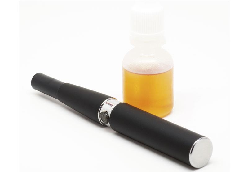
E‐cigarette vapour can “kill” bronchial epithelial cells


Use of electronic cigarettes (E-cigarettes) may result in bronchial epithelial apoptosis and macrophage efferocytosis dysfunction through the reduced expression of apoptotic cell recognition receptors, a study has shown. Additionally, nicotine alone appears to influence efferocytosis and some cytokines.
“E-cigarettes can cause airway epithelial cell death and apoptosis and efferocytic dysfunction of macrophages via alteration of apoptotic cell recognition receptors and can alter bronchial epithelial cytokine secretion pathways in a flavour-dependent manner with some variation between different apple E-liquids observed,” the researchers said.
“As such, E-cigarettes should be treated with caution by users, especially those who are nonsmokers,” they added.
An EVOD-2, which runs at 3.7 V and a resistance of 1.5 Ω, was used in all experiments in this study. Three apple flavours were tested from two suppliers in a 70-percent propylene glycol: 30-percent vegetable glycerine base, including one specially tested to confirm the absence of diacetyl (DA) and acetyl propionyl (AP).
Sytox Green stain and Annexin V were used to measure cell necrosis and apoptosis. Efferocytosis was measured by internalization of pHrodo Green labelled apoptotic airway cells by macrophages. Flow cytometry was then used to measure the expression of macrophage cell surface apoptotic cell receptors, and cytokine bead array to measure cytokine release by E-cigarette-exposed airway cells.
E-cigarette vapour led to an increase in primary bronchial epithelial necrosis and apoptosis, as well as reduced efferocytosis (lowest flavour, 12.1 percent) compared with control (20.2 percent; p=0.032). [Respirology 2020;25:620-628]
One flavour reduced the efferocytosis receptor CD44 (mean fluorescent intensity [MFI] vs control: 1,863 vs 2,332; p=0.016). All components reduced expression of CD36, including the glycol bases (MFI vs control: 1,067–12,274 vs 1,415). Of note, secretions of TNF‐α, IL‐6, IP‐10, MIP‐1α, and MIP‐1β were reduced for all flavour variants.
“Many studies now show that E-cigarettes have toxic effects on a range of cells, including airway epithelial cells, [but] many have been limited to looking at cell viability and looking at a range of flavours without investigating the variants in the same flavour,” the researchers said. [Respir Res 2016;17:57; Reprod Toxicol 2012;34:529-537; PLoS One 2015;10:e0118344; Clin Oral Investig 2016;20:477-483; J Environ Pathol Toxicol Oncol 2016;35:343-354; Toxicol Mech Methods 2016;26:427-434]
In this study, the E-cigarette vapour extract (EVE) from three apple E‐liquids ± nicotine (18 mg/mL) from two retailers were examined, including one free of DA‐AP, which has been associated with popcorn lung. [South Med J 2008;101:541-542; PLoS One 2013;8:e57935]
“We observed increased cytotoxicity with EVE exposure consistent with findings from other studies looking at the toxicity of EVE, many of which also show the effect is dependent on the flavour,” the researchers said. [PLoS One 2016;11:e0157337; Inhal Toxicol 2013;25:354-361; Oral Oncol 2016;52:58-65; Inhal Toxicol 2017;29:126-136; PLoS One 2015;10:e0116732; Thorax 2016;71:376-377]
In addition, the current findings showed that the release of DNA was flavour-specific and independent of nicotine, suggesting that despite the right chemoattractant being released, “the phagocytes will be unlikely to phagocytose their targets even if drawn to them,” according to the researchers.
“We look forward to reading future studies investigating damage-associated molecular pattern release,” they added.